

To the nose, and beyond
medical publications worth mentioning

rhinorrhea = runny nose
epistaxis = bloody nose
-
A thin cribiform plate:
Tim D. White, Pieter A. Folkens. “Skull” in The Human Bone Manual, 2005
“The cribriform plate roofs the nasal cavities, and because it is perforated by many tiny foramina it looks like a sieve. Olfactory nerves (cranial nerve 1) perforate this plate as they pass up to the brain from the mucous lining of the nose.”
Cribriform Plate Injury After Nasal Swab Testing for COVID-19
Knížek Z, Michálek R, Vodička J, Zdobinská P. 2021; doi:10.1001/jamaoto.2021.2216
“We present a case of cerebrospinal fluid (CSF) leak after skull base injury following nasal swab testing for COVID-19 in a patient with a previously intact skull base…
An otherwise healthy man in his 40s presented for right-sided clear water rhinorrhea in December of 2020. Rhinorrhea originated after nasal swab testing and was mistakenly considered to be allergic rhinitis in the patient. The test was performed by a mobile unit at the patient’s home in March of 2020. The test was indicated because of previous contact with a woman who had a positive COVID-19 test result 5 days earlier. The patient had no symptoms of COVID-19 infection and RNA of SARS-CoV-2 was not detected by polymerase chain reaction (PCR) testing. The patient had no other symptom except persistent unilateral nasal discharge from March to December 2020. …

Image: The yellow arrowhead points at the defect in cribriform plate on the right side of the nasal cavity. The dashed arrow represents the assumed trajectory of the nasal swab.
Clear nasal secretion … was noticeable during nasal endoscopy. On computed tomographic (CT) scan there was a defect in the lamina cribrosa on the right side (Figure). A previous brain CT scan from 2011 showed no skull base defect or other pathology. … Endonasal endoscopic closure was performed. … The patient reported anosmia … The score of an Odorized Markers Test was 8 points (hyposmia, testing both sides)….
A CSF fistula is a rare but dangerous complication. … Every instance of unilateral clear water rhinorrhea that appears after transnasal testing must be considered a potential CSF leak.”
-
-
CSF Leak After COVID-19 Nasopharyngeal Swab: A Case Report
2021: Paquin R, Ryan L, Vale FL, Rutkowski M, Byrd JK. doi: 10.1002/lary.29462.
The nasopharyngeal swab has been used with increased frequency since the beginning of the COVID-19 pandemic. Little has been written in the literature regarding the complications arising from this procedure, as it is generally accepted as safe. In this report, we describe a case in which a young woman sustained a traumatic skull base injury during a nasopharyngeal swab for COVID-19. … This case demonstrates the potential for significant complications arising from this widespread procedure and the necessity for awareness of these potential complications.
… The patient is a 38‐year‐old female who presented to our hospital after experiencing severe pain during an NP swab for COVID followed by 2 days of persistent clear watery rhinorrhea that worsened with leaning forward. She also noticed persistent headache and metallic tasting postnasal drip. The patient had no history of head trauma or surgery. She was otherwise healthy. … The patient underwent a computed tomography scan, which demonstrated a subtle defect in the posterior right cribriform plate (Fig. 1).

Image: Computed tomography scan without contrast in the (A) sagittal plane and (B) coronal plane demonstrating the defect in the right cribriform plate (arrows).
“The use of the NP swab has increased significantly since the beginning of the COVID‐19 pandemic. The NP swab has been utilized in the past, typically by experienced caregivers on hospitalized patients, to diagnose viral upper respiratory infections, but with the current public health strategy based on early detection and isolation, the test has been deployed on a massive scale …. The concern for CSF leak is significant, as 10%–25% of patients with traumatic CSF leaks will develop meningitis (Mourad et al. 2018).”
-
-
COVID‐19 swab‐related skull base injury
Sandeep G Mistry, Wallace Walker, James Earnshaw and Anders Cervin. 2021. doi: 10.5694/mja2.51082
“The patient precisely recalled the onset of unilateral clear rhinorrhoea, which occurred within hours of an “extremely painful” coronavirus disease 2019 (COVID‐19) swab test. … Intra‐operative evaluation confirmed a small, well demarcated defect (2–3 mm) in the left anterior skull base in the posterior cribiform plate…”
-
-
Olfactory fossa depth: CT analysis of 1200 patients
2018: Babu AC, Nair MRPB, Kuriakose AM. doi: 10.4103/ijri.IJRI_119_18.
“Olfactory fossa (OF) is a depression in anterior cranial cavity whose floor is formed by cribriform plate of ethmoid. Lateral lamella, which forms its lateral boundary, is a thin plate of bone and is at risk of injury during functional endoscopic sinus surgery, especially when fossa is deep/asymmetric.”
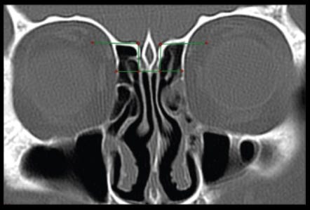
Image: Coronal CT demonstrating type III Keros classification. Here the olfactory fossae are very deep compared with types I and II.
[The round circles are the eyes.]
-
-
Identification of Significant Anatomical Variations in the Nose and Anterior Skull Base Using Computed Tomography: A Cross-Sectional Study
2020: Farhan N, Naqvi S, Rasheed B, et al. doi:10.7759/cureus.8449
… The identification of important anatomic variants of paranasal sinuses (PNS) is crucial in the planning of functional endoscopic sinus surgery (FESS) or other skull base surgical procedures…. Radiologists should identify and report these anatomical variations so that the operating surgeon anticipates technical challenges, and the patient can give informed consent [2].
…The Keros classification for olfactory groove defines depth in accordance with the lateral lamella cribriform plate height…”
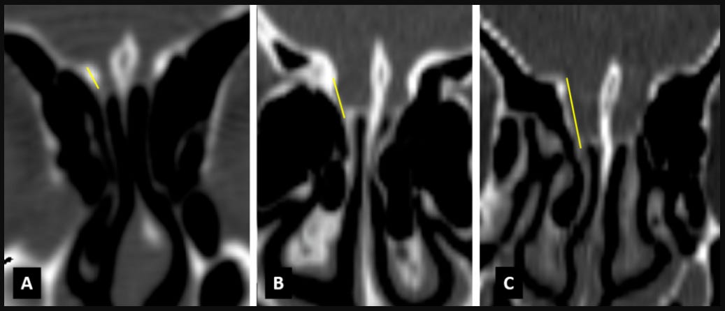
Figure 2: Coronal CT images showing Keros classification. Keros (A) type I bilaterally, (B) type II bilaterally, and (C) type III bilaterally.
-
-
The Preoperative Sinus CT: Avoiding a "CLOSE" Call with Surgical Complications.
2016: O'Brien WT Sr, Hamelin S, Weitzel EK. doi: 10.1148/radiol.2016152230.
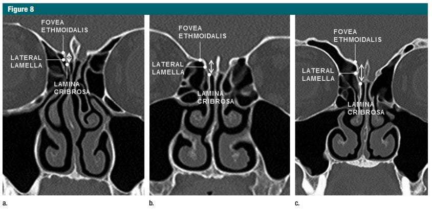
Image: …The anatomy of the cribriform plate/anterior skull base and varying depths of the olfactory fossae. The olfactory fossa is delineated by the horizontal lamina cribrosa [cribiform plate] inferiorly … Its depth is categorized according to the Keros classification (arrows).
…As the depth of the olfactory fossa increases, the lateral lamella becomes more vulnerable to intraoperative injury, either directly or through manipulation during turbinectomy or ethmoidectomy. Disruption of the lateral lamella results in direct communication between the paranasal sinuses and intracranial compartment, often with a cerebrospinal fluid (CSF) leak, which may be identified intraoperatively or present more insidiously in the postoperative setting. … Direct communication between the intracranial compartment and the sinus cavity substantially increases the risk of intracranial spread of infection and may also lead to development of a pseudomeningocele or meningoencephalocele.”
-
Performing nasopharyngeal swabs—Guidelines based on an anatomical study
2021: Pruidze P, Mincheva P, Weninger JT, Reissig LF, Hainfellner A, Weninger WJ. doi: 10.1002/ca.23762.
“…No systematic anatomical study defines concrete prerequisites for successfully targeting the nasopharyngeal mucosa. We therefore aim at simulating nasopharyngeal swabs in human body donors to characterize parameters allowing and supporting to enter the nasopharynx with a swab, while avoiding endangering the cribriform plate. … [Our data] demonstrate that the danger for damaging the cribriform plate or olfactory mucosa with swabs is unlikely, but potentially higher when performing nasal swabs.

Image: Required elevation of ala nasi. …(C, D) Probe inserted in the suggested direction along the hard palate. Note the very intense elevation of the ala nasi. C, anterior cranial fossa; N, nasal cavity; HP, hard palate; F, frontal bone; S, sphenoid bone; T, tongue; CV, bodies of cervical vertebrae.
[Intended technique: The swab must be pushing up on the nostrils, pointing slightly downward.]
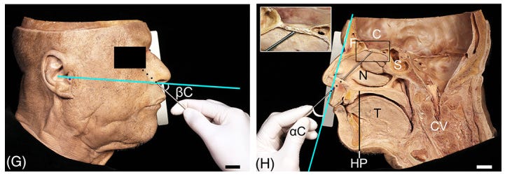
Image: Probe positions in respect to anatomical landmarks. … (G, H) Positioned to touch the cribriform plate [black box] …
[Wrong technique: if the swab is pointing upward, it is aiming for the cribiform plate.]
“… It is highly unlikely that the swab is pushed strongly enough to penetrate the cribriform plate. It is more likely that in such a case the swab causes an irritation of the olfactory mucosa and the olfactory nerve, which might result in temporary reduction of olfaction.
-
-
Anatomy by Reuters:

-
-
The Olfactory Epithelium and Olfactory Receptor Neurons
2001: Purves D, Augustine GJ, Fitzpatrick D, et al., editors. Neuroscience. 2nd edition. https://www.ncbi.nlm.nih.gov/books/NBK10896/
The transduction of olfactory information occurs in the olfactory epithelium, the sheet of neurons and supporting cells that lines approximately half of the nasal cavities. … The olfactory epithelium includes several distinct cell types (Figure 15.5A). The most important of these is the olfactory receptor neuron, a bipolar cell that gives rise to a small-diameter, unmyelinated axon at its basal surface that transmits olfactory information centrally. At its apical surface, the receptor neuron gives rise to a single process that expands into a knoblike protrusion from which several microvilli, called olfactory cilia, extend into a thick layer of mucus… that lines the nasal cavity …This entire apparatus—mucus layer and epithelium with neural and supporting cells—is called the nasal mucosa. The superficial location of the nasal mucosa allows the olfactory receptor neurons direct access to odorant molecules. Another consequence, however, is that these neurons are exceptionally exposed. Airborne pollutants, allergens, microorganisms, and other potentially harmful substances subject the olfactory receptor neurons to more or less continual damage.
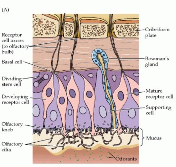
Image: Diagram of the olfactory epithelium showing the major cell types: olfactory receptor neurons and their cilia … Nerve bundles of unmyelinated neurons and blood vessels run in the basal part of the mucosa (After Anholt, 1987).
-
-
Anatomy by Reuters II
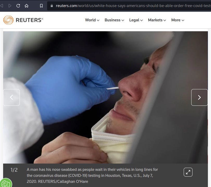
Image: July 7, 2020. REUTERS/Callaghan O'Hare. https://www.reuters.com/world/us/white-house-says-americans-should-be-able-order-free-covid-tests-later-this-2022-01-10/
-
-
[Best technique, risk-free and better outcome: saliva]
Saliva or Nasopharyngeal Swab Specimens for Detection of SARS-CoV-2
Wyllie AL, Fournier J, Casanovas-Massana A, et al. N Engl J Med. 2020;383(13):1283-1286. doi: 10.1056/NEJMc2016359
“We detected more SARS-CoV-2 RNA copies in the saliva specimens … than in the nasopharyngeal swab specimens …. In addition, a higher percentage of saliva samples than nasopharyngeal swab samples were positive.”
-
Reliability of induced sputum test is greater than that of throat swab test for detecting SARS-CoV-2 in patients with COVID-19: A multi-center cross-sectional study
Lai T, Xiang F, Zeng J, et al. Virulence. 2020;11(1):1394-1401. doi:10.1080/21505594.2020.1831342
“We previously reported that sputum induction was more sensitive than throat swabs for the detection of severe acute respiratory syndrome coronavirus 2 (SARS-CoV-2)… The positive rate for induced sputum was significantly higher than for throat swabs both overall (28.6% vs 5.4%, respectively; p < 0.01)…. Induced sputum is more reliable and has a lower false-negative rate than throat swabs…”
-
Cerebrospinal Fluid Leak From COVID-19 Swab
Agamawi YM, Namin A, Ducic Y. OTO Open. 2021 Nov 15;5(4):2473974X211059104. doi: 10.1177/2473974X211059104.
-
Meningitis due to cerebrospinal fluid leak after nasal swab testing for COVID-19
Alberola-Amores FJ, Valdeolivas-Urbelz E, Torregrosa-Ortiz M, Álvarez-Sauco M, Alom-Poveda J. Eur J Neurol. 2021 Nov;28(11):e91-e92. doi: 10.1111/ene.14736.
-
Misdirection of a nasopharyngeal SARS-CoV-2 swab: An unexpected complication
Cantarella G, Nava N, Pirondini C, Pignataro L. Otolaryngol Case Rep. 2022 Sep;24:100439. doi: 10.1016/j.xocr.2022.100439.
-
Iatrogenic rhinorrhea by nasal swab testing during COVID-19 pandemic: Case report
Demirci Otluoglu G, Paker B, Zorlu E, Akakın A, Kilic T. Neurochirurgie. 2022 Apr;68(3):347-348. doi: 10.1016/j.neuchi.2021.08.001.
-
Traumatic Cribriform Plate Defect Following Self-administered COVID-19 Nasal Swab Test
Douglas CF, White BD. Appl Radiol. 2021;50(4):44-46.
-
Covid-19 Nasopharyngeal Swab Related CSF Rhinorrhoea: A case report.
Dündar G, Özer S, Süslü AE, Önerci M. Indian J Otolaryngol Head Neck Surg. 2022 Mar 26:1-3. doi: 10.1007/s12070-022-03096-z.
-
Is oro/nasopharyngeal swab for SARS-CoV-2 detection a safe procedure? Complications observed among a case series of 4876 consecutive swabs
2021: Fabbris C, Cestaro W, Menegaldo A, Spinato G, Frezza D, Vijendren A, Borsetto D, Boscolo-Rizzo P. doi: 10.1016/j.amjoto.2020.102758.
-
Cerebrospinal fluid leak following a COVID-19 nasopharyngeal swab
Hill T, Sivapatham S, Metcalfe C, Tzortzis S. Br J Hosp Med (Lond). 2021 Dec 2;82(12):1-3. doi: 10.12968/hmed.2021.0529.
-
COVID-19 nasopharyngeal swab causing a traumatic cerebrospinal fluid leak
Ovenden C, Bulshara V, Patel S, Vsykocil E, Valentine R, Psaltis A, Abou-Hamden A. ANZ J Surg. 2021 May;91(5):1021-1022. doi: 10.1111/ans.16910.
-
Rhinorrhea following SARS-CoV-2 nasopharyngeal swab: A case for β2-transferrin testing
Perneczky J, Neuchrist C, Sellner J. Eur J Neurol. 2021 Nov;28(11):3552-3553. doi: 10.1111/ene.14883.
-
CSF rhinorrhoea post COVID-19 swab: A case report and review of literature
Rajah J, Lee J. J Clin Neurosci. 2021 Apr;86:6-9. doi: 10.1016/j.jocn.2021.01.003.
-
CSF Leak Following Nasal Swab Testing For COVID-19
Sadashiva A, Panji N, Shivappa L. Neurol India. 2021 Sep-Oct;69(5):1467-1468. doi: 10.4103/0028-3886.329562.
-
CSF rhinorrhea after nasopharyngeal swab testing for COVID-19: A case report and review of literature
Samadian M, Maroufi SF, Taheri MS, Jafari A. Otolaryngol Case Rep. 2021 Nov;21:100370. doi: 10.1016/j.xocr.2021.100370.
-
Cerebrospinal Fluid Leak After Nasal Swab Testing for Coronavirus Disease 2019
Sullivan CB, Schwalje AT, Jensen M, Li L, Dlouhy BJ, Greenlee JD, Walsh JE. JAMA Otolaryngol Head Neck Surg. 2020 Dec 1;146(12):1179-1181. doi: 10.1001/jamaoto.2020.3579.
-
A brief report: Cerebrospinal fluid rhinorrhea after repetitive nasal swab testing for coronavirus disease 2019(COVID-19)
Yılmaz M, Bahadır Z, Madendere B, Yüksel RT, Gökay H, Yiğitbaşı AA. Otolaryngol Case Rep. 2021 Sep;20:100313. doi: 10.1016/j.xocr.2021.100313.
-
Les prélèvements nasopharyngés ne sont pas sans risque
Bull Acad Natl Med. 2021 Jun;205(6):555-556. French. doi: 10.1016/j.banm.2021.04.013.
Oh for the love of cheese. Are you freaking KIDDING me? So are you saying inserting six inch long stick into the nose is NOT good? Are you saying the extreme discomfort caused by doing that could be a sign it should be avoided? Oh the number of times I was told not to “be a baby” …
Seriously, my family has avoided those weird tests the best we can but have still had a few each just for the honor of conducting our lives … I am pretty mad at this point.