Intramyocardial Inflammation after COVID-19 Vaccination: An Endomyocardial Biopsy-Proven Case Series
Baumeier, Christian, Ganna Aleshcheva, Dominik Harms, Ulrich Gross, Christian Hamm, Birgit Assmus, Ralf Westenfeld, Malte Kelm, Spyros Rammos, Philip Wenzel, Thomas Münzel, Albrecht Elsässer, Mudather Gailani, Christian Perings, Alae Bourakkadi, Markus Flesch, Tibor Kempf, Johann Bauersachs, Felicitas Escher, and Heinz-Peter Schultheiss. 2022.
https://www.mdpi.com/1422-0067/23/13/6940 https://doi.org/10.3390/ijms23136940
-
"Myocarditis in response to COVID-19 vaccination has been reported since early 2021. In particular, young male individuals have been identified to exhibit an increased risk of myocardial inflammation following the administration of mRNA-based vaccines. Even though the first epidemiological analyses and numerous case reports investigated potential relationships, endomyocardial biopsy (EMB)-proven cases are limited. Here, we present a comprehensive histopathological analysis of EMBs from 15 patients with reduced ejection fraction (LVEF = 30 (14–39)%) and the clinical suspicion of myocarditis following vaccination with Comirnaty® (Pfizer-BioNTech) (n = 11), Vaxzevria® (AstraZenica) (n = 2) and Janssen® (Johnson & Johnson) (n = 2). Immunohistochemical EMB analyses reveal myocardial inflammation in 14 of 15 patients, with the histopathological diagnosis of active myocarditis according the Dallas criteria (n = 2), severe giant cell myocarditis (n = 2) and inflammatory cardiomyopathy (n = 10). Importantly, infectious causes have been excluded in all patients. The SARS-CoV-2 spike protein has been detected sparsely on cardiomyocytes of nine patients, and differential analysis of inflammatory markers such as CD4+ and CD8+ T cells suggests that the inflammatory response triggered by the vaccine may be of autoimmunological origin. Although a definitive causal relationship between COVID-19 vaccination and the occurrence of myocardial inflammation cannot be demonstrated in this study, data suggest a temporal connection. The expression of SARS-CoV-2 spike protein within the heart and the dominance of CD4+ lymphocytic infiltrates indicate an autoimmunological response to the vaccination."

Image: Evidence of SARS-CoV-2 spike protein in cardiac tissue after COVID-19 vaccination. (A–C) Representative immunohistochemical stainings of SARS-CoV-2 spike protein in EMBs from patients diagnosed with DCMi after receiving Comirnaty® (panel A and B, patients 5 and 10) or Vaxzevria® (panel C, patient 13). (D) SARS-CoV-2-positive cardiac tissue served as positive control.
-
-
-
Co-expression of the SARS-CoV-2 entry molecules ACE2 and TMPRSS2 in human ovaries: Identification of cell types and trends with age
Wu M. et al., 2021. doi: 10.1016/j.ygeno.2021.08.012. PMID: 34418496
“… In this study, public datasets containing bulk and single-cell RNA-Seq data derived from ovarian tissues were analyzed to demonstrate the mRNA expression and protein distribution of the two key entry receptors for SARS-CoV-2—angiotensin-converting enzyme 2 (ACE2) and type II transmembrane serine protease (TMPRSS2)…. Co-expression of ACE2 and TMPRSS2 was observed mostly in oocytes and partially in granulosa cells….
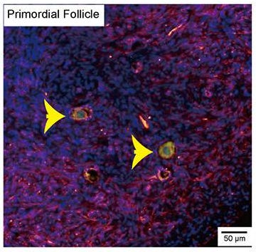

Images: Immunofluorescence staining showed the co-expression of ACE2 and TMPRSS2 in human ovaries. The yellow arrows indicate follicles. The blue channel is DAPI staining, the green channel is ACE2 expression, and the red channel is TMPRSS2 expression.
… Bian et al. [2020, PMID: 34192086] reported that SARS-CoV-2 RNA and viral particles were detected in multiple organs and tissues including the testis and ovary, which indicated that SARS-Cov-2 can reach the reproductive organs. Pathological analysis of the testes displayed varying degrees of spermatogenic cell reduction and damage. Using public datasets, Wang and colleagues [PMID: 32283711] also provided evidence that the human testis are a potential target site of SARS-CoV-2 infection. The ovary is the core of the female reproductive system, and damage to it can cause infertility and premature ovary failure….
… Our study revealed that approximately 80% of the ovarian cells were positive for ACE2 and TMPRSS2; they are predominantly enriched in oocytes and matrix cells of human ovaries. Additionally, there was no observed variation in ACE2 and TMPRSS2 expression in ovaries of different ages…”
-
-
SARS-CoV-2 Infection of Human Ovarian Cells: A Potential Negative Impact on Female Fertility
2022: Luongo FP, Dragoni F, Boccuto A, Paccagnini E, Gentile M, Canosi T, Morgante G, Luddi A, Zazzi M, Vicenti I, Piomboni P. doi: 10.3390/cells11091431. PMCID: PMC9105548
“… Here, we investigated the potential of SARS-CoV-2 to infect the follicular microenvironment, in particular granulosa (GCs) and cumulus cells (CCs), thus providing evidence for a productive infection. GCs and CCs were recovered from women (n = 25) who underwent in vitro fertilization at the Assisted Reproductive Unit, Siena University Hospital. … We demonstrated the expression of cell host factors ACE2, TRPMSS2, BSG and CTSL, which are pivotal for the virus life cycle. Cultured GCs and CCs incubated with SARS-CoV-2 revealed productive SARS-CoV-2 infection at 24 h, 48 h and 72 h post-adsorption. Indeed, SARS-CoV-2 RNA, spike and nucleocapsid proteins were detected in GCs and CCs, and their cell culture supernatant successfully infected the standard VERO E6 cells. Finally, TEM showed full-size virions attached to the membrane and located inside the cytoplasm. This in vitro study reveals the susceptibility of human ovarian cells to SARS-CoV-2 infection, suggesting a potential detrimental effect of COVID-19 infection on female human fertility.”
-
-
Hidden Picture: See if you can find the cardiomyocytes or oocytes in this informative illustration below, courtesy of New York Times.
-
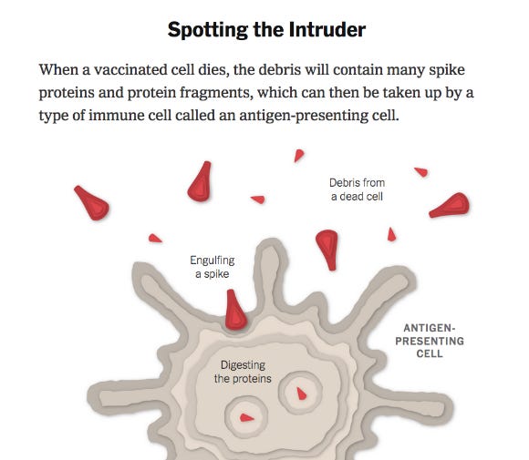
Spotting the Intruder
“When a vaccinated cell dies, the debris will contain many spike proteins and protein fragments...” (New York Times, Corum and Zimmer. “How Moderna’s Vaccine Works”. May 7, 2021)